Home » Uncategories » Back Of Neck Anatomy - Anatomy of The Neck: Causes of Neck Pain and How to Manage ... - Use the mouse scroll wheel to move the images up and down alternatively use the tiny arrows (>>) on both side of the image to move the images.
Back Of Neck Anatomy - Anatomy of The Neck: Causes of Neck Pain and How to Manage ... - Use the mouse scroll wheel to move the images up and down alternatively use the tiny arrows (>>) on both side of the image to move the images.
ads/wkwkland.txt
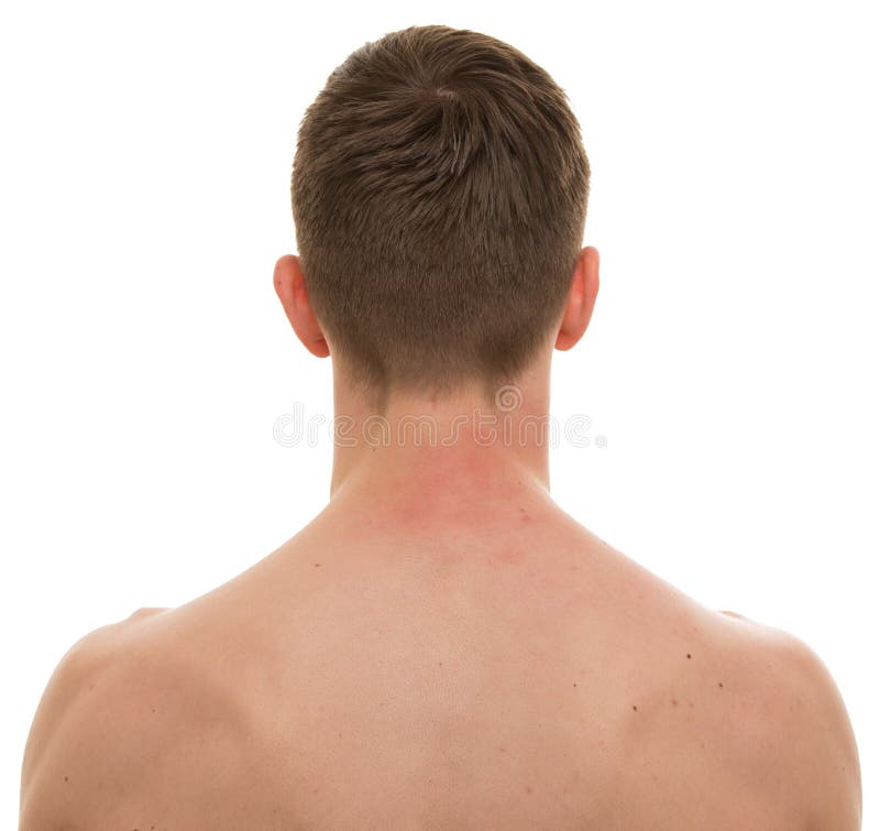
Back Of Neck Anatomy - Anatomy of The Neck: Causes of Neck Pain and How to Manage ... - Use the mouse scroll wheel to move the images up and down alternatively use the tiny arrows (>>) on both side of the image to move the images.. D) demonstrate sound knowledge of the surface/living and radiological anatomy. Muscles of the posterior neck and the back. Neck muscle anatomy body anatomy anatomy drawing anatomy art figure drawing reference anatomy reference human muscular system human anatomy for artists muscles of the neck. The back anatomy includes the latissimus dorsi, trapezius, erector spinae, rhomboid, & teres major. The anterior muscles of the neck facilitate swallowing and speech.
Of the head, neck and vertebral column; Last time we learned that the trapezius makes the back wall of the neck. Our neck is where we find the seven cervical vertebrae, with c7 (the seventh cervical vertebra) meeting t1 (the first thoracic vertebra) at the base of the neck. This article describes the anatomy of the head and neck of the human body, including the brain, bones, muscles, blood vessels, nerves, glands, nose, mouth, teeth, tongue, and throat. It runs from the neck to the upper back.
Male Neck Back Isolated On White - REAL Anatomy Stock ... from thumbs.dreamstime.com The back anatomy includes the latissimus dorsi, trapezius, erector spinae, rhomboid, & teres major. See more ideas about anatomy, anatomy and physiology, muscle anatomy. D) demonstrate sound knowledge of the surface/living and radiological anatomy. Your neck is like no other part of the vertebral spinal column and enables your head and neck a wide range of motion. We've largely focused on the physical aspect of our spinal anatomy in this series. Learn vocabulary, terms and more with flashcards, games and other study tools. Muscles of the posterior neck and the back. From the sides and the back of the neck, the splenius capitis inserts onto the head region, and the splenius cervicis extends onto the cervical region.
Learn vocabulary, terms and more with flashcards, games and other study tools.
Learn about these muscles, their locations & functional the traps are quite a complex set of muscles. Integrates anatomy and physiology of cells, tissues, organs, the systems of the human body, and mechanisms responsible for homeostasis. It is made up of bones discs muscles ligaments nerves and tendons. Learn vocabulary, terms and more with flashcards, games and other study tools. The structure is, of course, an important part of the conversation. 3d video tutorials and interactive modules on the anatomy of the back including anatomy of the musculature, vertebral column, joints and ligaments. Discography is a diagnostic procedure the back experts at the southeastern spine institute (ssi) use to determine if any of your intervertebral discs are the primary cause of your back pain. A collection of anatomy notes covering the key anatomy concepts that medical students need to learn. The cervical spine supports the weight and movement of your head and pro. Some important structures contained in or passing through the neck include the seven cervical vertebrae and enclosed spinal cord, the jugular veins and carotid arteries, part of the esophagus, the larynx. Posterior border of the ligament is free, anterior border is attached to the cervical spines and its superior border. They control the scapulae (shoulder blades), which play a role in shrugging, neck movement, head. Beneath the integument the back of neck presents in the median plane the ligamentum nuchae, which is a triangular fibrous sheet and represents upward continuation of supraspinous ligament.
Anatomical principles underlying cranial nerve lesions; The splenius muscles originate at the midline and run laterally and superiorly to their insertions. Beneath the integument the back of neck presents in the median plane the ligamentum nuchae, which is a triangular fibrous sheet and represents upward continuation of supraspinous ligament. Neck, in land vertebrates, the portion of the body joining the head to the shoulders and chest. Of the head, neck and vertebral column;
Male Neck Back Isolated On White - REAL Anatomy Stock ... from thumbs.dreamstime.com « back show on map ». Despite being a relatively small region, it contains a range of important anatomical features. The sections below will cover these elements in more detail. The cervical spine supports the weight and movement of your head and pro. Your neck is like no other part of the vertebral spinal column and enables your head and neck a wide range of motion. From the sides and the back of the neck, the splenius capitis inserts onto the head region, and the splenius cervicis extends onto the cervical region. Many in the neck help to stabilize or move the head. They control the scapulae (shoulder blades), which play a role in shrugging, neck movement, head.
Discography is a diagnostic procedure the back experts at the southeastern spine institute (ssi) use to determine if any of your intervertebral discs are the primary cause of your back pain.
Your neck is like no other part of the vertebral spinal column and enables your head and neck a wide range of motion. The splenius muscles originate at the midline and run laterally and superiorly to their insertions. The head rests on the top part of the vertebral column, with the skull joining at c1. We've largely focused on the physical aspect of our spinal anatomy in this series. Muscles of the posterior neck and the back. Want to learn more about it? 3d video tutorials and interactive modules on the anatomy of the back including anatomy of the musculature, vertebral column, joints and ligaments. Head and neck anatomy is important when considering pathology affecting the same area. Top head neck anatomy flashcards ranked by quality. The suprahyoid muscles originate from above the hyoid bone in the chin region. Start studying anatomy of neck & back. This article describes the anatomy of the head and neck of the human body, including the brain, bones, muscles, blood vessels, nerves, glands, nose, mouth, teeth, tongue, and throat. The spine runs from the base of your skull down the length of your back, going all the way down to your pelvis.
Some important structures contained in or passing through the neck include the seven cervical vertebrae and enclosed spinal cord, the jugular veins and carotid arteries, part of the esophagus, the larynx. Clinically, surface anatomy is used to split the neck into anterior and posterior triangles which provide clues as to the location of specific structures. Click now to study the muscles, glands and organs of the neck at kenhub! Many in the neck help to stabilize or move the head. The sections below will cover these elements in more detail.
Nerves of the Head and Neck | Interactive Anatomy Guide from www.innerbody.com The back anatomy includes the latissimus dorsi, trapezius, erector spinae, rhomboid, & teres major. It provides images in the axial and coronal planes so that the user can study and learn anatomy. Discography is a diagnostic procedure the back experts at the southeastern spine institute (ssi) use to determine if any of your intervertebral discs are the primary cause of your back pain. The neck is the area between the skull base and the clavicles. Many in the neck help to stabilize or move the head. A collection of anatomy notes covering the key anatomy concepts that medical students need to learn. The head rests on the top part of the vertebral column, with the skull joining at c1. This is often a result of incorrect posture.
E) demonstrate practical lab skills in anatomy and an appreciation of the ethics.
Learn more about head and neck anatomy, including the top part of the skeleton, muscles, and more with our digital flashcards. Sternocleidomastoid muscle (main muscle in the front of the neck) thyroid gland From the sides and the back of the neck, the splenius capitis inserts onto the head region, and the splenius cervicis extends onto the cervical region. The spine runs from the base of your skull down the length of your back, going all the way down to your pelvis. Despite being a relatively small region, it contains a range of important anatomical features. The sections below will cover these elements in more detail. This is often a result of incorrect posture. Posterior border of the ligament is free, anterior border is attached to the cervical spines and its superior border. In radiology, the 'head and neck' refers to all the anatomical structures in this region excluding the central nervous system, that is, the brain and spinal co. The levator scapulae muscle is attached at the top four cervical vertebrae (c1 to c4) and runs down the side of the neck to attach at the top of the shoulder blade (scapula). It is made up of bones discs muscles ligaments nerves and tendons. Discography is a diagnostic procedure the back experts at the southeastern spine institute (ssi) use to determine if any of your intervertebral discs are the primary cause of your back pain. The suprahyoid muscles originate from above the hyoid bone in the chin region.
ads/wkwkland.txt
0 Response to "Back Of Neck Anatomy - Anatomy of The Neck: Causes of Neck Pain and How to Manage ... - Use the mouse scroll wheel to move the images up and down alternatively use the tiny arrows (>>) on both side of the image to move the images."


0 Response to "Back Of Neck Anatomy - Anatomy of The Neck: Causes of Neck Pain and How to Manage ... - Use the mouse scroll wheel to move the images up and down alternatively use the tiny arrows (>>) on both side of the image to move the images."
Đăng nhận xét